
Διαγνωστικές & θεραπευτικές τεχνικές στις παθήσεις του θώρακα


Διαγνωστικές & θεραπευτικές τεχνικές στις παθήσεις του θώρακα

Evangelos Perdikakis & Vasilios Skiadas
Introduction
Given the fact that magnetic resonance imaging (MRI) is being performed more frequently for assessment of the knee joint (e.g. post-traumatic, in sport injuries, in rheumatological disorders, in oncological imaging), the number of incidental cystic and “cyst-like” lesions in and around the knee joint found on routine knee MRI scans has also increased [1–4]. The vast majority of these lesions are benign, ranging from benign cysts to complications of underlying diseases and many of them demonstrate characteristic features on MRI, thus allowing a confident diagnosis to be made [1–6]. Knowledge of the common anatomical locations and appearances of bursae, recesses, cysts and ganglia is necessary so that radiologists do not misinterpret these benign entities as soft-tissue tumours [1–8]. It is of paramount importance for the radiologist to be aware of the MRI features because understanding the spectrum of appearances of the various benign cystic lesions is vital for optimal patient management. This article is intended to be a comprehensive pictorial review of the most common and uncommon benign cystic and “cyst-like” lesions in and around the knee joint. For easier classification purposes, benign cysts were subdivided into categories as following: (1) synovial cysts, (2) ganglion cysts, (3) meniscal cysts and (4) intraosseous cysts. Similarly, “cyst-like” lesions were subclassified into the following: (1) normal knee bursae, (2) normal knee recesses and (3) miscellaneous cyst-like lesions.
Evangelos Perdikakis, Evangelia G. Chryssou, Apostolos Karantanas
University of Crete, Heraklion, Greece
Introduction
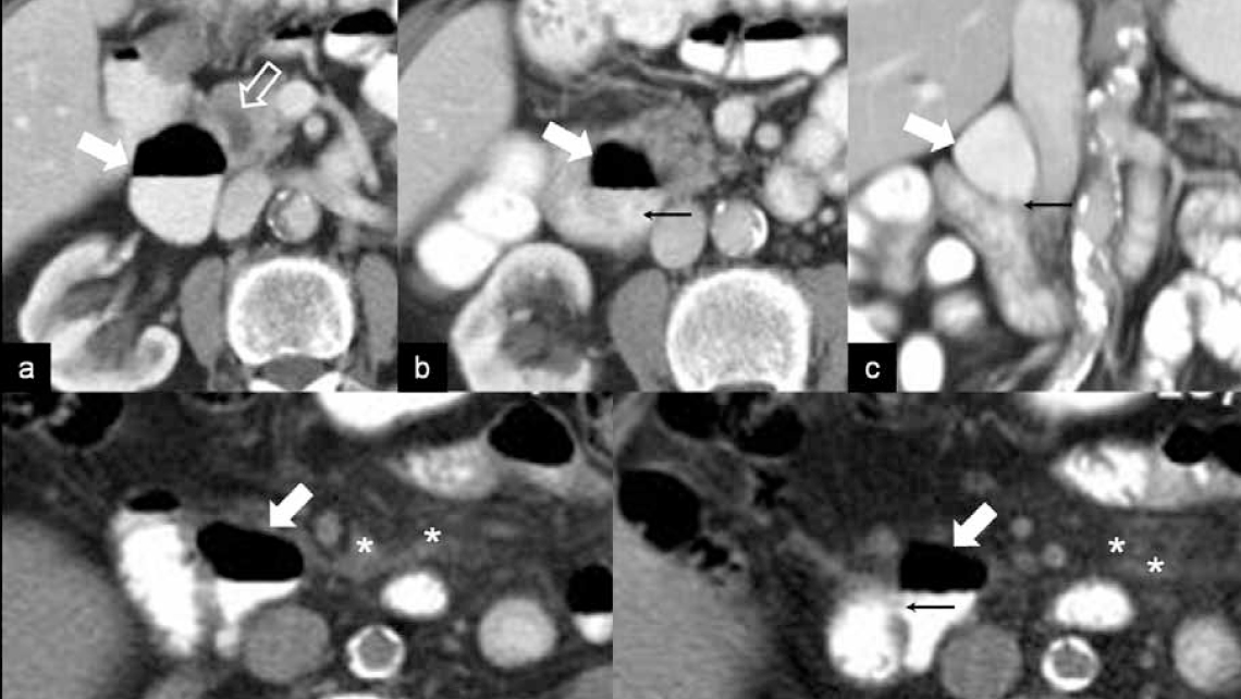
Duodenal diverticula occur very commonly with a reported incidence of 1- 6% in upper gastrointestinal barium studies and prevalence as high as 22% in autopsy studies [1,2]. Despite their incidence, duodenal diverticula are usually asymptomatic [2]. For that reason the duodenum is often overlooked on cross sectional imaging studies as an underlying cause in the setting of acute abdominal symptomatology [3]. Due to the continuing evolution of Multidetector Computed Tomography (MDCT) and Magnetic Resonance Imaging (MRI) technology and the application of post-processing techniques, accurate depiction and demonstration of duodenal anatomy and pathology is now feasible [4,5]. Although the role of imaging in complications resulting from duodenal diverticula has been described, to the best of our knowledge this is the first study focusing solely on acute abdominal symptomatology. Furthermore we provide comparative data regarding the depiction of duodenal periampullary diverticula in the axial and coronal plane in MDCT exams and secondly we examine the ability of diverticular neck demonstration in the aforementioned planes.

Αγαπητέ επισκέπτη,
Η Επεμβατική Ακτινολογία είναι η κλινική υποειδικότητα της Ακτινολογίας, που με την χρήση μηχανημάτων τελευταίας τεχνολογίας μπορούμε να εφαρμόσουμε σύγχρονες διαγνωστικές και θεραπευτικές τεχνικές με τον ελάχιστα επεμβατικό τρόπο. Είναι σημαντικό για τους ασθενείς και τους θεράποντες ιατρούς τους, να είναι επαρκώς πληροφορημένοι για όλες τις δυνατές επιλογές που τους προσφέρει η σύγχρονη ελάχιστα επεμβατική ιατρική και σκοπός της ιστοσελίδας μου είναι να σας ενημερώνω με ακρίβεια και με υπευθυνότητα για τις διαγνωστικές τεχνικές και θεραπείες που προσωπικά εφαρμόζω, παρουσιάζοντας κάποια ενδεικτικά αποτελέσματα από περιστατικά ασθενών μου.
Ευάγγελος Ν. Περδικάκης
![]()


